Echocardiogram (Echo)
Often referred to as echo, is a non-invasive imaging technique that uses ultrasound waves to create real-time images of the heart's structure and function. It is commonly used to evaluate the heart's chambers, valves, blood flow, and surrounding structures to diagnose and monitor various heart conditions.
Indications
- Valvular Heart Disease (e.g. mitral regurgitation, aortic stenosis, etc.)
- Congenital Heart Defects (e.g. atrial septal defect, ventricular septal defect, etc.)
- Heart Failure - to assess the heart's pumping funtion and ejection fraction (the percentage of blood the left ventricle pumps out with each contraction)
- Cardiomyopathy - to evaluate heart muscle function, whether it's dilated, hypertrophic, or restrictive.
- Aneurysms - to check for dilation in the heart's arteries, such as an aortic aneurysms.
- Endocarditis - to identify infections of the heart valves.
- Arrhythmias - to evaluate heart rhythm and detect abnormal patterns or electrical disturbances.
- Heart Murmurs - to determine if they are caused by a structural issue in the heart (like a valve problem).
- Pericardial Effusion - to check for fluid around the heart which can be caused by infection, inflammation or injury.
How the procedure works
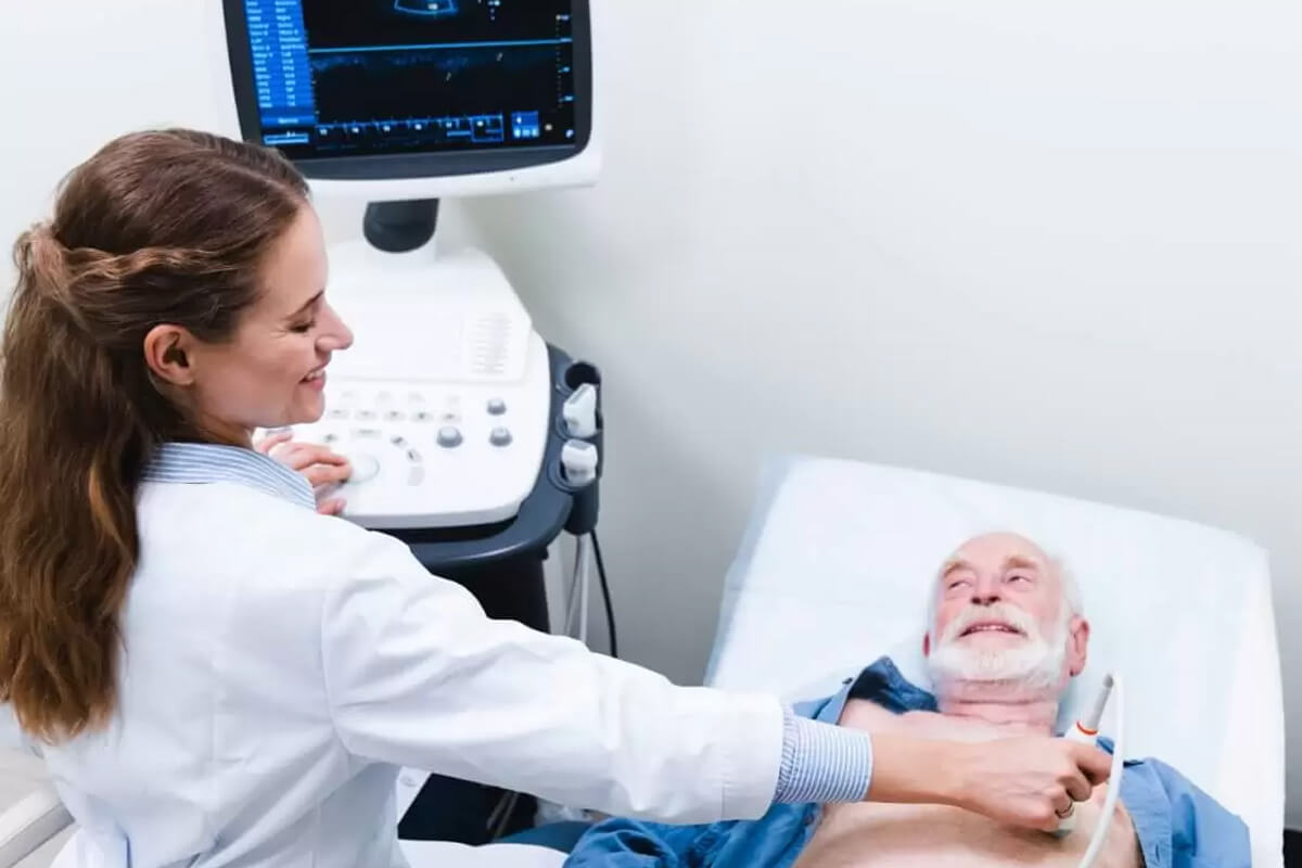
Preparation: In most cases, there is no special preparation for a standard echocardiogram. You may be asked to remove clothing from the upper body and wear a gown.
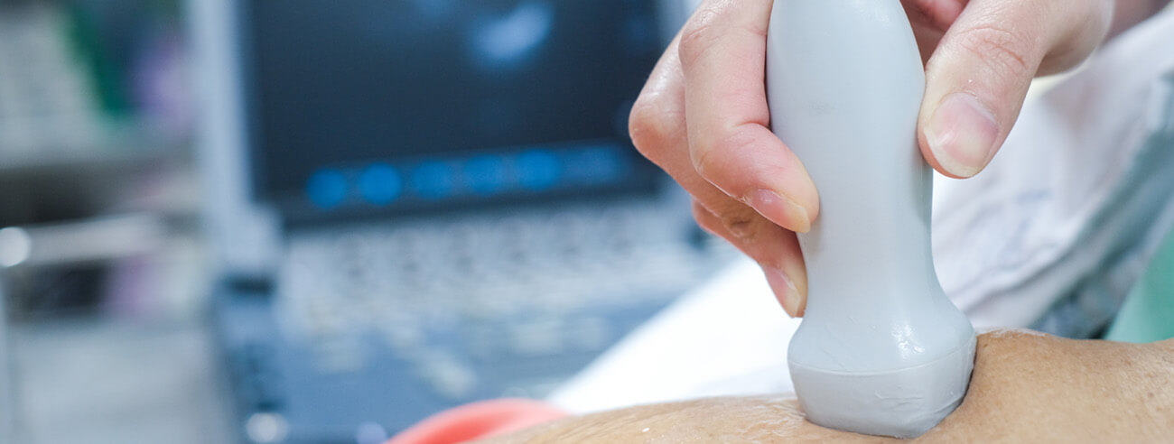
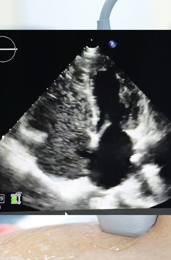
Procedure: You lie on an exam table, usually on your left side, while the technician applies a gel to your chest to improve transmission of sound waves. The transducer (probe) is moved over the chest to capture images from different angles. You may be asked to breathe deeply or change position during the exam to obtain better images.
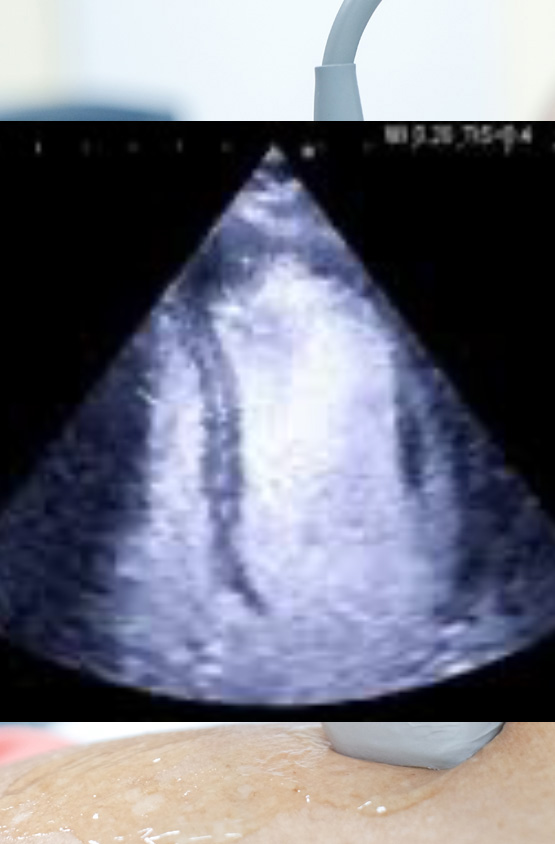
During the procedure: The technician uses the images from the echocardiogram to evaluate heart structures and function. The test may take about 30 to 60 minutes for routine and longer for specialized techniques. Specialized techniques include but are not limited to:In some cases, an ultrasound enhancer called Definity may be used to optimize the images. A bubble study may be ordered for certain indication like a stroke or TIA. Cardia strain analysis is often performed for oncology patients and for certain cardiac conditions such as Amyloidosis.
Heart wall motion abnormalities - Identifying parts of the heart muscle that aren't moving properly, often due to a heart attack. Valve problems - Identifying valve stenosis or regurgitation (leakage). Heart enlargement - Enlarged chambers or thickened heart muscle. Blood Clot - Detection of Infection or Inflammation - Detection of infections in the heart lining or surrounding sac.Non-invasive - Unlike CT scans or MRIs, echocardiography doesn't involve radiation.
Real-Time Imaging - Provides live images of the heart's movement, allowing doctors to assess its function in real time.
Echocardiograms can be limited by poor image quality in patients with obesity or difficulty lying in position.
After your echocardiogram, you may resume all normal activities - there are no restrictions.
Conclusion
Echocardiography is a valuable tool in the diagnosis and management of a wide range of heart conditions. It is often the first test used to assess heart function, and its non-invasive nature makes it safe and accessible for many patients. If any abnormalities are found, your healthcare provider will discuss the next steps for treatment or further testing.
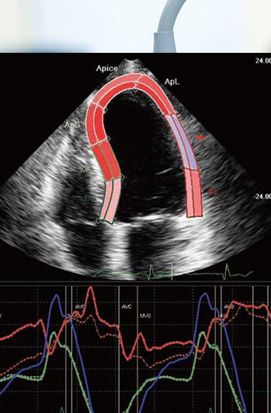
Measures the deformation of the heart's muscular tissue.
Detect wall motion abnormalities when the images are suboptimal.
Visualize blood flow through the heart to detect any adnormal connections.