Upper Extremity Arterial Duplex
An Upper Extremity Arterial Duplex is a non-invasive ultrasound test that evaluates the arteries in the arms. It combines traditional ultrasound imaging with Doppler ultrasound to assess both the structure of the arteries and blood flow patterns. An Upper Extremity Arterial Duplex is an essential diagnostic tool for evaluating and managing vascular health in the arms, ensuring early detection and treatment of potentially serious conditions.
Purpose of an Upper Extremity Arterial Duplex:
- Diagnose arterial disease in the arms, such as: Artherosclerosis (plaque buildup causing narrowing or blockages), Blood clots (thrombosis) or embolisms, Aneurysms (buldging of the arterial wall).
- Identify vascular trauma or injuries.
- Assess Raynaud's phenomenon, a condition causing reduced blood flow to the fingers.
- Monitor blood flow after vascular surgeries, such as Bypass Grafts of Stents.
Indications for the Test:
- Pain or cramping in the arms during use.
- Non-healing wounds or ulcers on the hands or arms.
- Symptoms of cold, pale, or discolored extremities.
- Suspected thoracic outlet syndrome (compression of blood vessels or nerves).
- A history of vascular conditions or surgeries in the arms.
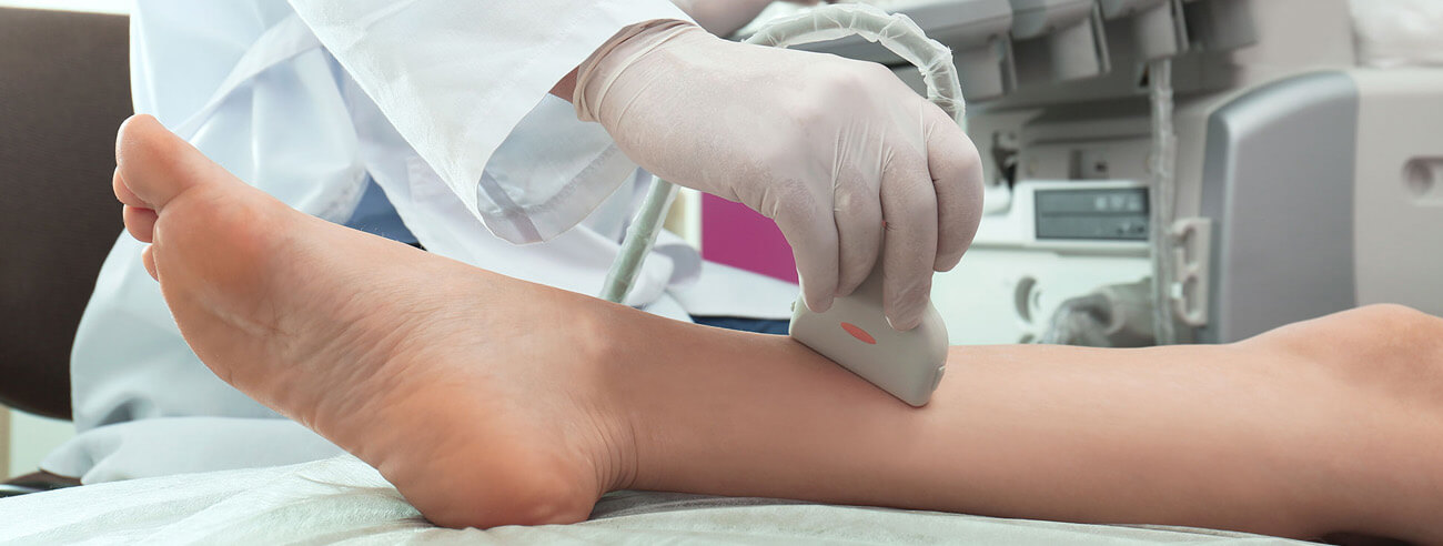
Procedure:
No specific preparation is required for this procedure, and patients are advised to wear loose-fitting clothing to allow easy access to their arms. During the examination, the patient either lies down or sits comfortably on an examination table with their arms extended. A water-based gel is applied to the skin over the arteries in the arms to ensure proper contact with the ultrasound probe. The probe is then gently moved along the arm to capture detailed images of the arteries. The Doppler component of the ultrasound measures blood flow velocity and direction, helping to identify any abnormalities such as blockages or reduced circulation. The entire procedure is non-invasive, painless, and typically takes between 30 to 60 minutes to complete.
Results:
Normal findings from an arterial ultrasound of the arms indicate that the arteries are open, free of blockages, and demonstrate normal blood flow patterns. Blood flows smoothly without any signs of turbulence or obstruction. Abnormal findings, however, may reveal narrowing of the arteries caused by plaque buildup (atherosclerosis), blood clots blocking blood flow, or the presence of an aneurysm, which is a bulging or weakened section of an artery. Additionally, vascular compression may be detected, often associated with conditions such as thoracic outlet syndrome, where nearby structures compress the arteries and affect circulation. Identifying these abnormalities is crucial for determining appropriate treatment and preventing complications.